3. Anatomical organization of the brain
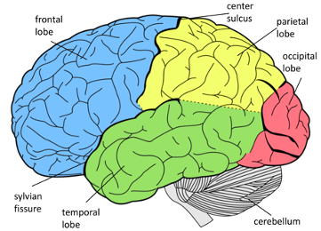
The cerebrum is the bulkiest part of the brain. It is described as the organ of thought and serves as the site of control of the nervous system; it allows the human being to possess the qualities associated with consciousness, such as perception, communication, understanding and memory (Kandel et al. 2013). The cerebral hemispheres consist of elevations (gyrus) and valleys (grooves); a longitudinal cerebral fissure separates the hemispheres that are kept connected through a set of fibers that constitute the corpus callosum. Each cerebral hemisphere is divided into lobes, which correspond, broadly speaking, to the overlying bones of the skull.
Frontal lobe. The frontal lobe is located in the anterior cranial fossa. The central sulcus divides the frontal lobe from the parietal lobe into a coronary plane. The gyrus anterior to the central sulcus is called the precentral sulcus and serves as the primary motor area of the brain. The rest of the frontal lobe is used in the modification of motor actions. It is in charge of deciding the appropriate motor behavior in each case. For example, when we place our hand differently to take a cup or a spoon, the way the hand acts is defined and decided in the frontal lobe. In the frontal lobe there is a band of tissue that acts as an anatomical map of our body, “the motor homunculus”, where the size of each body segment is proportional to the complexity of the movement and action to be performed. Thus, the hand, fingers and face have, on this map, a greater extension than the shoulders or hips.
The frontal lobe is very developed in the human being hosting important non-motor tasks such as behavior planning, control of our emotions, reasoning, and judgment, which are complex functions not always easy to analyze if a patient has a stroke. The areas responsible for these skills are ahead of those devoted to motor function (premotor and prefrontal areas).
Parietal lobe. The parietal lobe interprets sensations coming from the body. The gyrus posterior to the central sulcus, the postcentral sulcus, is the primary area for the reception of these sensations. To act, we need information from our environment and our own body. Thus, continuing with the example of the cup of coffee, we could not perform such a simple task, if we did not detect through our senses the weight of the teaspoon we move, the size of it, the map of where our hand is and the route we must take. The parietal lobe is involved in the map of “where to act”, integrating sensory information interoceptive (from our body: muscles, joints, tendons) and exteroceptive (from the outside). It is basically attributed sensory, associative functions, as well as recognition of space.
Occipital lobe. The occipital lobe is located above the tent of the cerebellum, in the posterior cranial fossa, and is mainly related to vision. It elaborates visual information, although it transcends the parietal and temporal lobes
Temporal lobe. The temporal lobe is located in the middle cranial fossa and is mainly related to hearing, as well as the place where, on its medial face, important structures of memory (hippocampus) and the unconscious emotional system (limbic system) sit.
In humans, a lateral shift of functions is recognized, so that the two cerebral hemispheres do not do the same. Evolution has been responsible for making the most of the brain through a division of labor between its two halves (the cerebral hemispheres). Thus, the right hemisphere focuses on nonverbal expression, perception and spatial orientation, and also on emotional behavior. Then, the right hemisphere thinks and remembers in images. Several studies have shown that people with a dominant right hemisphere study, think, remember and learn in images, as if it were a film without sound. These people are very creative and have very developed imaginations.
On the contrary, the left hemisphere (more developed in most human beings) is responsible for verbal development, which contributes to its production and understanding. In it there are two structures that are closely related to linguistic ability, the “Broca Area” and “Wernicke’s Area” (areas specialized in language and exclusive to the human being). The specific function of the “Broca Area” is oral expression, it is the area that produces speech. The “Wernicke Area” has as its specific function the understanding of language since it is the receptive area of speech.
In addition to the verbal function, the left hemisphere has other functions, such as the ability to analyze, ability to make logical reasoning, abstractions, solve numerical problems, learn theoretical information, make deductions, etc.
But the brain, despite dividing the work between different regions and hemispheres, functions as a unit, achieving in real time a coordinated and precise action. Below the cortical mantle (cerebral cortex) is the cerebral white matter through which bundles of nerve fibers cross, each with a different course and type of information. Below this white matter are located the deep gray nuclei (basal ganglia) that are involved in multiple functions, especially in motor behavior.
Brain development and cognitive maturation occur concurrently during childhood and adolescence (Casey et al. 2005). Brain regions associated with more basic functions such as motor and sensory processes mature first, followed by association areas involved in top-down control of thoughts and action. The total brain size is about 90% of adult size by age 6 years, the brain continues to undergo dynamic changes throughout adolescence and well into young adulthood.
At the fourth month of gestation, a differentiation of cells is observed in the fetus; similarly, neurons and glial cells are produced at an important rate. Through a migration process, these cells form the first brain regions that will ensure the most elementary functions, such as reflex movement, physical behaviors, and balance. The areas in charge of sensory stimuli, memory, and emotions are formed a little later: the cerebral cortex, which is the part of the brain that is associated with higher cognitive activities such as attention, synthesis, planning, reasoning, spatial imagination, and language.
While it is true that during childhood several important changes in mental abilities and brain maturation are noticed, the adolescent brain continues its development, even beyond adolescence and ends up reaching its maximum volume around the age of twenty-five. Even after reaching this peak the brain does not lose its plasticity. This plasticity is also manifested in the ability of certain cortical regions to assume functions that, in principle, would be carried out by regions that have lost their functionality, as a result of relatively minor damage.
If you want to know more about the parts of the human brain, as well as its unique defenses, like the blood-brain barrier, watch the next video:
For further information about the anatomical organization of the brain, check the additional resources:
- Kolb & Whishaw (2019). Introduction to brain and behavior.
- Gazzaniga, M. S., Ivry, R. B., & Mangun, G. R. (2019). Cognitive neuroscience: The biology of the mind.
- Ward (2020). The Student’s Guide to Cognitive Neuroscience.